Intrasense
Advanced visualization solutions for medical imaging
Description
Intrasense, a French expert in medical imaging since 2004, develops and markets in 40 countries unique medical devices, brought together in software platforms that facilitate diagnosis, decision making and patient follow-up.
Myrian®, an advanced visualization solution for radiology, optimizes image interpretation using expert tools assisted by Artificial Intelligence. A multidisciplinary and collaborative platform dedicated to oncology is currently being developed, which will make patient management and therapeutic follow-up more fluid.
The design of these innovative solutions is possible thanks to the daily work of a team of 66 enthusiasts, an investment of more than 12 million euros in Research & Development since its creation and a close collaboration with health professionals, to offer them tools focused on their needs.
Contact
Featured products
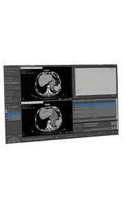
Cardiovascular system
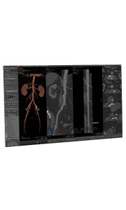
XP-Vessel
Myrian XP-Vessel permits to visualize a complete vessel analysis. Anomalies are detected more quickly, which makes diagnosis more secure.
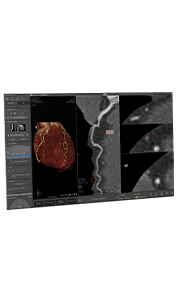
XP-Cardiac
Myrian XP-Cardiac simplifies the analysis and diagnosis of CT cardiovascular exams with dedicated cardiac coronary pathology analysis tools.
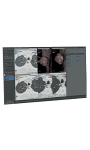
Oncology
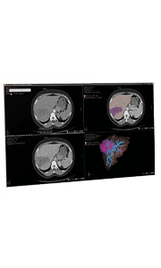
XP-Liver
XP-Liver, a clinical application dedicated to the detection of liver lesions, helps segment the liver and its lesions, especially for surgery.
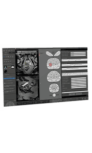
XP-Prostate
XP-Prostate, focused on prostate MRI examinations, accelerates the creation of reports thanks to advanced and automated post-processing tools.
XP-Onco
XP-Onco provides innovative tools for analysis, interpretation, diagnosis and evaluation of therapeutic response, dedicated to oncology follow-up.
Lungs
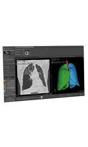
XP-Lung
XP-Lung allows the precise analysis of a lung examination (segmentation, analysis of the lung parenchyma and airways) to simplify the diagnosis.
XP-Lung Nodule
XP-Lung Nodule is an advanced clinical application for the analysis of cancerous lesions and the segmentation, quantification and follow-up of lung nodules.